Escherichia coli
Escherichia coli, or E. coli also called coli, an intestinal bacterium is very common among mammals humans. Discovered in 1885 by Theodor Escherich in stools of infants, is usually a commensal fecal coliform. However, some strains of E. coli can be pathogenic thus inducing gastroenteritis, urinary tract infections, meningitis, or sepsis.
Description
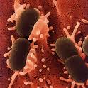
E. coli is a gram-negative bacillus radiation-immune family Enterobacteriaceae femtograms weighing 110. It is a common host of commensal intestinal microflora of man and warm-blooded animals (mammals and birds). Its establishment in the digestive tract occurs during the first hours or days after birth. E. coli is so throughout the life of the host bacterial species dominant aerobic intestinal flora. E. coli is probably the most studied organism on this day since the age of its discovery and easily cultivated (cell division every 20 minutes at 37 ° C in a rich medium) make it a tool study of choice. The profusion of scientific publications that mention it witnessed, and she plays the role of "workhorse" in all molecular biology laboratories.
History
Theodor Escherich, observing the frequency of neonatal diarrhea, had already raised the issue of involvement in the E. coli enteritis. After World War II, knowledge has converged to establish the concept of virulence of certain strains of E. coli. In the 1950s, many strains of E. coli have been implicated as causative agent of infantile diarrhea. We now know that certain strains "specialized" E. coli are associated with very different pathologies (including extra-intestinal), both in humans and in animals, diarrhea, gastroenteritis, urinary tract infections, meningitis, septicemia, "hamburger disease", the hemolytic Uremic etc..
For prevention, surveillance of HUS at the Centre National de Reference E. coli, located in the Biodiversity Unit of Emerging Bacterial Pathogens to the Pasteur Institute (France), which is responsible for studying the pathogenic strains.
Since the 1950s, bacteriologists have tried, through the antigenic differences of E. coli, to subdivide the species into serotypes by immunizing rabbits with somatic and flagellar antigens. The serogrouping remains the method most used today.
The serotype is the combination of the two antigens, somatic O and flagellar H (eg O157: H7 and O111: H8), while serogroup is determined by the O antigen (eg, O157, O111). However, the serotype is not sufficient to characterize the E. coli pathogens. Each serotype is not necessarily correlated with pathogenicity.
Recently, a laboratory in California (LS9) explained that this bacterium uses to produce hydrocarbons, a technique that may be responsible for the synthesis of oil.
Antigens and serogrouping
The somatic O antigen defining serogroup, is contained in the lipopolysaccharide present on the cell wall of Gram-negative strains. H flagellar antigen is proteinaceous structure within the flagellum (cilliature peritrichous) allowing the mobility of the bacteria. The K antigen surface is not always present but if present, it blocks the agglutinability of the antigen O.
The somatic O antigens
There are over 150. The somatic antigens are composed of complex lipopolysaccharide. Currently some medical laboratories using agglutination with sera to determine the serogroup, but this technique is limited by the number of higher and higher serum to produce by the presence of cross-agglutination between the O antigens of E . coli, Shigella, and those of Salmonella, and the passage of the creamy consistency of the colony textured with the consequent lack of synthesis of O antigen For this reason, a molecular serotyping technique has been developed.
The O antigen is part of the lipopolysaccharide (LPS) of the outer membrane of Gram-negative bacteria. It contains a large number of repeating units of oligosaccharides from March to June sugars whose combination determines the diversity of antigens O. The genes encoding enzymes involved in the synthesis of O antigen are clustered in the rfb gene cluster.
The rfb cluster can be amplified specifically with a primer and, after restriction endonuclease MboII, a profile rated "R" can be obtained by electrophoresis, corresponding to serogroup E. coli. A profile of electrophoresis is based on the location of restriction sites suitable for MboII. Thus all clusters of genes corresponding to a somatic antigen have a restriction pattern of its own. This profile is then analyzed with R software Taxotron ® and compared to a database, in perpetual development. For example, the profile will have a number R111 R, corresponding to serogroup O111 obtained with serum.
Flagellar H antigens
The H antigens are not used to identify E. coli pathogens but have a great interest in epidemiological perspective: the identity of the H antigen is a component to ensure that it is the same strain.
The diversity of antigen H is due to different types of flagellin component structure of the flagellum. It allows the flagellum motility. Typing is also achieved by agglutination, but is developed in very few laboratories in the world. However, some strains lose their mobility and are classified as non-mobile (NM or M-). A molecular serotyping technique has also been developed to determine the antigen H.
The H antigen is encoded by the gene fliC. The N and C terminal parts of flagellin are highly conserved and is the middle part, more variable, which gives the specificity of antigen H. The E. coli also still have the fliC gene but are unable to synthesize functional flagella. After amplification and restriction of the fliC gene, it is possible to type the H antigen by comparing the pattern obtained from a database of profile-type. For example, the profile fliC (rated E) has a number F8, corresponding to the type H8 obtained with serum.
The surface antigen or envelope K
There are three types of K antigen designated by the letters L, A or B.
* The antigen is the most common but is heat labile (it was destroyed in half an hour. 100 ° C). So the heating causes a loss of antigenicity, the power to fix the antibodies and the power to hide the O antigen
* The A antigen is rare, it is a capsular antigen (encapsulated E. coli are relatively frequent in urinary tract infections). The Hep A is highly thermostable (you have to destroy autoclaving)
* The B antigen is always present in E. enthéropathogènes coli of infantile gastroenteritis. It has a thermolability intermediate: after half an hour. 100 ° C, there is always B antigen O antigen but may come into contact with the serum by "holing" of the envelope, antibody binding is always positive but the antigenicity is lost gradually (depending on the duration of heating).
Difference between antigen and B antigen A or L: in a homogeneous culture dish,
* 80% + of colonies and 20% of colonies - for A or L
* Homogeneous distribution throughout the population for B.
Read also Appendicitis
wikipedia
